Collimation Effects
X-ray beam collimation for radiography and fluoroscopy projection imaging is important for patient dose and image quality reasons. Actively collimating to the volume of interest reduces the overall integral dose to the patient and thus minimizes the radiation risk. Less volume irradiated will result in less x-ray scatter incident on the detector. This results in improved subject contrast and image quality.
X-ray field collimation differs from the use of electronic magnification in that the acquired field of view remains constant, and there is no improvement in the resultant spatial resolution performance (see below). However, the use of collimation will normally reduce the image brightness, and require a corresponding increase in radiation entrance skin dose to the patient, although not to the level when electronic magnification is used, because the minification gain is unchanged.
Pelvis phantom
 |
 |
 |
| Figure Q | Figure R | Figure S |
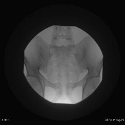
The three images shown above (Q, R, and S) show the effect of collimating the x-ray beam whilst maintaining a constant 38 cm diameter input field of view. As one collimates the x-ray beam in going from Figure Q to Figure S, less of the patient is exposed, but the image characteristics of the central region are essentially unchanged. In particular, there is no improvement in spatial resolution that can be achieved by the use of electronic zoom where the acquired field of view is electronically reduced (see above). The image in Figure Q used 77 kV/2.5 mA which resulted in an entrance air kerma rate of 39 mGy/minute. By contrast, the image in Figure R used 79 kV/2.6 mA which resulted in an entrance air kerma rate of 40 mGy/minute and the image in Figure S used 84 kV/2.7 mA and resulted in 46 mGy/minute.
The use of collimation generally increases the entrance air kerma rate, which is a very important consideration if there is any possibility of inducing deterministic effects such as epilation and erythema. However, the threshold dose for deterministic effects is conservatively taken to be at least ~2 Gy, and this value is only likely to be reached in interventional radiology. For most fluoroscopy examinations deterministic effects are not expected, and the patient radiation risk is proportional to the total energy imparted to the patient. The stochastic radiation risk is therefore proportional to the product of the entrance exposure air kerma rate and the exposed area. If the exposed area is halved, the corresponding increase in entrance air kerma rate will be less than a factor of two because of the increase in x-ray tube voltage. Accordingly, provided there is no risk of the induction of deterministic radiation effects, increased collimation during fluoroscopy should reduce the risk of patient stochastic effects, and is therefore strongly recommended.
The use of collimation in fluoroscopy does not significantly affect the overall image quality in terms of spatial resolution or scatter when the II input field of view is unchanged. The spatial resolution performance, which is inversely proportional to the input field of view, is constant (note that the display field of view does not change in Figures Q, R, and S). The amount of scatter is not expected to significantly change from the reduction in the total exposed patient mass; fluoroscopy is performed with the use of scatter removal grids that eliminate 90% or more of the scattered radiation. Any reduction in scatter in Figure S relative to Figure O would likely be far too small to be perceptible for most clinical applications.
Skull phantom
 |
 |
| Figure T | Figure U |
Figure T shows a frame from a non-collimated fluoroscopy run obtained using a 25 cm field of view. The radiographic techniques for this image were 74 kV/2.2 mA, and the corresponding entrance air kerma rate was 26 mGy/minute. Note the brightness at the edge of the image where x-ray beam directly impacts on the image intensifier, which will reduce the display contrast of the anatomical features of interest. Figure Q shows the improvement that is achieved in terms of display contrast by the use of collimation. In Figure U, the radiographic techniques used were 83 kV/2.6 mA, which resulted in an entrance air kerma rate of 40 mGy/minute. In this example, considerations of image quality are paramount, and the use of collimation is strongly recommended because of the marked improvement in the resultant display contrast (i.e., display contrast is not "wasted" to depict the air around the patient).
