Auto Modes
Auto-Filter (Anthropomorphic phantom)
Figure A shows an image acquired using the anthropomorphic breast phantom with the digital mammography system operating in Auto Filter mode. The image shown in Figure A was acquired using a Rhodium filter, and an x-ray tube voltage of 32 kV. The system terminated the exposure at 40 mAs, and the recorded Exposure Index (EI) value was 464. The system also indicated an entrance air kerma of 6.6 mGy, which resulted in an average glandular dose 0.88 mGy.
It is important to note that the phantom material composition caused the digital mammography to use a very penetrating beam of radiation. However, the fact that the phantom is relatively compact results in an artificially low average glandular dose. It is therefore very important to focus on the relative doses to the anthropomorphic phantom, since the phantom composition does not permit the absolute doses to be interpreted in a meaningful manner.
 |
| Figure A. Image of the anthropomorphic phantom obtained using the auto filter mode of operation, which automatically selects both the x-ray beam filter, as well as the optimum x-ray tube voltage. Click the image for a half-size version, View the full size image. |
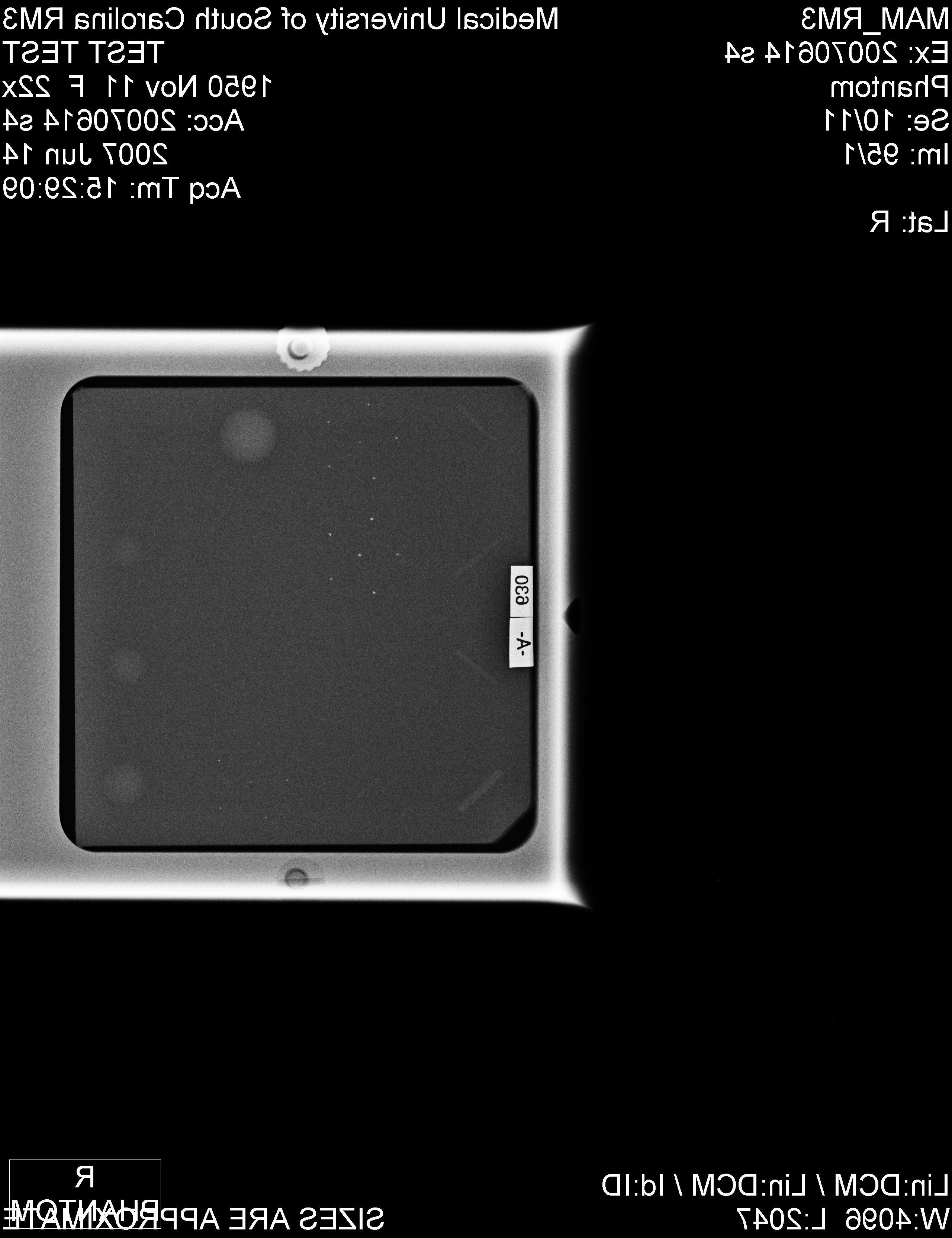
Auto-Filter (ACR Accreditation Phantom)
Figure B shows an image acquired using the ACR accreditation image quality phantom with the digital mammography system operating in auto filter mode. The image shown in Figure B was acquired using a Molybdenum filter, and an x-ray tube voltage of 29 kV. The system terminated the exposure at 53 mAs and the recorded Exposure Index (EI) value was 440. The system also indicated an entrance air kerma of 8.8 mGy mGy, which resulted in an average glandular dose 1.5 mGy. Note that the phantom material composition caused the digital mammography to use a much less penetrating beam of radiation than in the example shown in Figure A. In the example shown in Figure A, the average glandular dose is close to the target that was selected for this imaging system by the manufacturer.
 |
| Figure B. Image of the anthropomorphic phantom obtained using the auto filter mode of operation, which automatically selects both the x-ray beam filter, as well as the optimum x-ray tube voltage. Click the image for a half-size version, or View the full size image. |
Auto kV
Figure C shows images acquired using the Auto kV mode, but using different x-ray beam filters that were manually selected by the operator. The left image in Figure C is of the ACR accreditation image quality phantom acquired using the Mo filter. For this image, the system selected 29 kV and terminated the exam after a total exposure of 53 mAs and an EI value of 440, which is very similar to the exposures depicted in Figure B above (Auto Filter mode). The entrance air kerma was 8.8 mGy and the average glandular dose was 1.5 mGy.
The right hand image in Figure C shows the corresponding acquisition made when the operator pre-selected the use of a Rhodium filter, but permitted the digital mammography system to select the x-ray tube voltage that it considered to be optimal. In this example, the x-ray tube voltage selected by the system was 31 kV, and the exposure was terminated at 40 mAs corresponding to an exposure index of 440, which is the same value that was reached when the Mo filter was selected. For the Rh/31 kV exposure shown in Figure C (right), the incident air kerma was 5.6 mGy, and the corresponding average glandular dose was 0.97 mGy.
This example illustrates the following features of the use of an auto kV mode, with the filter choice manually selected by the operator:
A. The exposure index value should remain constant, irrespective of the actual filter or kV choice.
B. Increasing the filter atomic number (i.e., choosing Rh), and increasing the voltage from 29 to 31 kV, increases the x-ray beam photon energy.
C. Increased photon energy will generally reduce the mAs; in the example in Figure C from 53 mAs to 40 mAs, which generally results in a shorter exposure time (i.e., less motion blur).
D. Increased photon energy will generally reduce the average glandular dose; in the example in Figure C from 1.5 mGy to 0.97 mGy.
 |
 |
| Figure C. Images obtained of the ACR accreditation image quality phantom obtained in auto kV and with the choice of x-ray beam filter material performed by the operator: Left Mo filter ( View the full size image ); Right Rh filter ( View the full size image ). Click each image for a half-size version. |