Anthropomorphic phantom
One important characteristic of any digital imaging system is to vary the amount of radiation used to generate any image. The gray scale appearance of digital images, unlike those of analog screen-film systems, is not affected by how much radiation was used. Accordingly, unlike screen-film systems, digital images can never be "under-exposed" (i.e., all white) or "over-exposed" (i.e., all black). When radiation intensity increases, quantum mottle will decrease and vice versa.
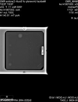
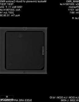
The two images depicted in Figure F were obtained using a Molybdenum x-ray beam filter and an x-ray tube voltage of 28 kV. The Left image in Figure F used 40 mAs. The entrance air kerma was 6.5 mGy, and the resultant average glandular dose was 0.69 mGy. The right image in Figure G shows the effect of further reducing of the radiation intensity, where the radiation used was only 4.7 mAs, and entrance air kerma of 0.80 mGy, and an average glandular dose of 0.077 mGy. As expected, when the filter and x-ray tube voltage are kept constant, there is a direct and simple proportionality between the mAs, entrance air kerma, and the average glandular dose. Figure G shows region of interest that illustrate the marked increase in quantum mottle as the amount of radiation that is used to generate these images is progressively reduced.
 |
 |
| Figure F. Images acquired of the anthropomorphic phantom in manual mode using identical exposure conditions (i.e., Mo/28 kV) except the total amount of radiation used to generate the image: Left 40 mAs ( View the full size image ); Right 4.7 mAs/EI 90 ( View the full size image ). |
| Figure G. Regions of interest of the anthropomorphic phantom images acquired in manual mode using identical exposure conditions (i.e., Mo/28 kV) except the total amount of radiation used to generate the image: Left 40 mAs; Right 4.7 mAs. |
ACR accreditation phantom
Images depicted in Figure H were obtained in the manual mode using a Molybdenum x-ray beam filter and an x-ray tube voltage of 25 kV. The Left image in Figure H used 110 mAs. The entrance air kerma was 13 mGy, and the resultant average glandular dose was 1.7 mGy. The right image in Figure H shows the effect of reducing of the radiation intensity, where the radiation used was only 26 mAs, or a less than a quarter of the value used for the image on the left. The image shown on the right in Figure H had an entrance air kerma of 2.7 mGy, and an average glandular dose of 0.34 mGy. Figure I shows region of interest that illustrate the marked increase in quantum mottle as the amount of radiation that is used to generate these images is progressively reduced, and illustrate how the visibility of lesions is reduced when less radiation is used to generate any digital image because of the increase in quantum mottle.
 |
 |
| Figure H. Images acquired of the ACR accreditation image quality phantom in manual mode using identical conditions (i.e., Mo/28 kV) except the total amount of radiation used to generate the image: Left 110 mAs ( View the full size image ); Right 26 mAs ( View the full size image ). |
| Figure I. Region of interest of the images shown in Figure H, and acquired in manual mode using identical conditions (i.e., Mo/28 kV) except the total amount of radiation used to generate the image: Left 110 mAs; Right 26 mAs. |
Digital versus Screen-Film
Figure J shows an image of the ACR accreditation phantom acquired on a digital imaging system using the techniques normally used in screen-film mammography (i.e., Mo filter + 25 kV), but using the auto timed mode of operation to ensure that the detector dose is the one normally used for this system (i.e., EI = 450). The total exposure used was 110 mAs; the entrance air kerma is 13 mGy, and the average glandular dose is 1.7 mGy. This image can be directly compared with Figure B, where the auto filter mode also selected the Molybdenum x-ray beam filtration, but a higher x-ray tube voltage (29 kV vs 25 kV). In the example shown in Figure J, use of a lower x-ray tube voltage (i.e., lower photon energy) will result in a higher average glandular dose (at 25 kV the dose is 1.7 mGy whereas at 29 kV the dose is 1.5 mGy).
Digital systems generally use higher photon energies (kV) than analog screen-film. Digital data can compensate loss of subject contrast at higher kVs by increasing the image display contrast. Accordingly, digital images use higher x-ray beam qualities because these reduce patient doses, and can compensate for the corresponding reduction in subject contrast by increasing the image display contrast. National surveys show that the introduction of digital mammography has resulted in a corresponding downward trend in radiation doses. At the same time, the ability to digitally improve (i.e., enhance) mammograms has also appeared to improve diagnostic imaging performance of this imaging modality.
| Figure J. Image acquired of the ACR accreditation phantom using manually selected values of filter (i.e. Mo) and x-ray tube voltage (i.e., 25 kV) that are normally used in screen-film mammography. The resultant image used 110 mAs, generated an exposure index of 450, and resulted in an average glandular dose of 1.7 mGy ( View the full size image ). |


